CHAPTER 1Introduction
Endochondral ossification is the process in which growth plates propagatelongitudinally and link the metaphysical and epiphyseal region inside the bone.Endochondral ossification comprises proliferation of chondrocytes, production ofextracellular matrix (ECM), hypertrophy of chondrocytes, calcification of matrix,vascular invasion, degradation of matrix and finally bone formation (Orth and Cook,1994). Longitudinal growth in long bones starts where resting chondrocytes ofepiphyseal growth plate differentiate into proliferative chondrocytes and form columns offlattened cells and these cells differentiate into hypertrophic zone of epiphyseal growthplate (Kronenberg, 2003).Long bone contains three major parts: (1) epiphysis is the rounded part of a longbone contain secondary ossification center; (2) metaphysis is the wide part of long bonelocated between epiphysis and diaphysis contain growth plates; and (3) Diaphysis is themain shaft of long bone contain primary ossification center (Boskey, 1985; Olsson andEkman, 1982; von Pfeil and Decamp, 2009) (Figure 1.1).
1.1 Long bone development
Bone growth is a highly coordinated process, any deviation from the normal trackmay lead to bone dysplasia. The growth of the bones is accomplished by the cartilagearea of the epiphyseal growth plate located at both ends of the long bone. Epiphysealgrowth plate (GP) is composed of chondrocyte and extracellular matrix (ECM), mainlyresponsible for the synthesis and elongation of long bones. ECM is mainly composed ofcollagen, proteoglycan, elastin and cartilage adhesive proteins. Chondrocytes within GParranged along the long axis is divided into different regions. The expression ofchondrocytes in different regions was different in morphology, differentiation, secretion,intracellular enzyme activity, growth factor and hormone receptor (Farquharson and Jefferies, 2000). Through the transport of nutrients in the blood vessels, the chondrocytesin the GP can proliferate, differentiate and secrete matrix proteins in a precise and orderlymanner, and finally, complete the bone growth (Hall et al., 2006).
..........
1.2 Growth plate cartilage
The growth plate cartilage is mainly made up of chondrocytes and responsible foractual bone length and growth rate. Growth plate can be divided into several differentzones on the basis of morphology (Figure 1.2 and Figure 1.3).Chondrocytes in this zone are flat and arranged in a column shape, and the secretionability of matrix protein was enhanced. In proximal part of proliferative zone, actual celldivision starts along with Collagen II production(Farquharson et al., 1992).This zonecontain highest volume of ECM and aggrecan(Alini et al., 1992).Transition zone also known as avascular zone, located between the proliferative andprehypertrophic zone. Growth plates receive blood from two types of blood vessels namedas proximal and metaphyseal. These two blood vessels do not meet and in this way createthe avascular zone or transition zone in the growth plate (Howlett et al., 1984; Hunt et al.,1979; Leach and Monsonego-Ornan, 2007)
..........
CHAPTER 2The effect of inhibition of hypoxia inducible factor-1α in thiraminduced tibial dyschondroplasia
2.1 Objectives
To identify the genotypic expression of HIF-1α and Hsp90 and their protein levelin avian growth plate before and after treatment. To evaluate the efficacy of synthetic medicine FK228 as HIF-1α inhibitor inthiram induced avian TD. To check association between HIF-1α and Hsp90 in thiram induced TD.
.........
2.2 Materials and Methods
All animal experiments were planned according to the national legislations foranimal welfare and following the approval and strict guidelines of the Institution AnimalCare and Use Committee of Huazhong Agricultural University Wuhan, China.Total 150 day old broiler chicks were bought from commercial hatchery (Chia TaiAnimal Husbandry Co. Ltd, Wuhan, China) and raised under standard temperature andhygienic conditions. Initially the chicks were divided into two groups, a control group(n=50) and a thiram group (n=100). Both groups were fed with standard normal diet andthiram group received in addition of tetramethylthiuramdisulphide (thiram) at the rate of50 mg/kg for the induction of disease according to (Shahzad et al., 2014a) after four dayspost-hatch. From day 8, half of birds were separated from the thiram group and allocatedas FK228 group and administer at the rate of 0.5 mg/kg/day intraperitoneally and normalsaline was administered to all the control birds. The dose of FK228 was designated basedon studies using FK228 to prevent tumor development in mice (Lee et al., 2003). All ofthree groups were raised for 14 days and number of lame birds was recorded in each groupon daily basis. Twenty-five birds from each group were euthanized by cervical dislocationon day 10 and day 14. The cartilage growth plate of some tibial bones were dissected outand were fixed in 4% paraformaldehyde and some of the bones were immediately frozen inliquid nitrogen and stored at 70°C for further analysis. TD was scored according to Pineset al. (2005).
...........
CHAPTER 3 Recovery of chicken growth plate by Hsp90 Inhibitor EGCG........23
3.1 Objectives .....23
3.2 Materials and Methods ............23
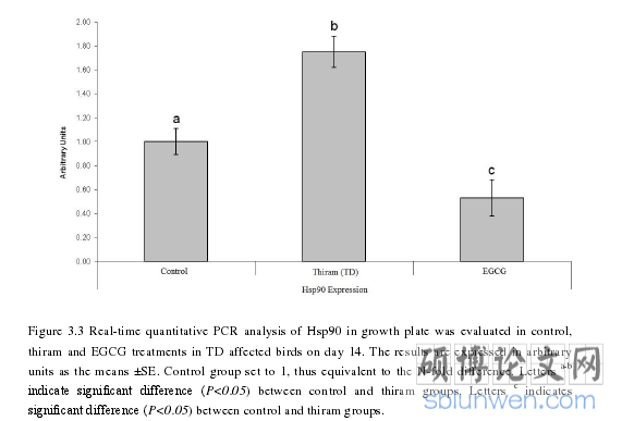
3.3 Results...........25
3.3.1 Effect of EGCG on thiram-induced TD-affected growth plates ......25
3.3.2 Effect of Apigenin on thiram-induced TD-affected growth plate ....28
3.4 Discussion.....31
CHAPTER 4 Therapeutic effect of hypoxia inducible factor-1α inhibitor ..... 33
4.1 Objectives..... 33
4.2 Material and Methods .... 33
4.2.1 Statistical analysis.... 34
4.3 Results .......... 35
4.4 Discussion .... 37
CHAPTER 5 Conclusions and Future prospects .... 40
5.1 Study I .......... 40
5.2 Study II......... 40
5.3 Study III ....... 40
5.4 Future prospects .... 41
CHAPTER 4Therapeutic effect of hypoxia inducible factor-1α inhibitorApigenin on liver toxicity in thiram induced tibialdyschondroplasia
4.1 Objectives
To study the effects of HIF-1α inhibition by Apigenin for the control andtreatment of avian TD. To analyze serum biomarkers and antioxidant liver enzymes in thiram induce TDbefore and after Apigenin treatment. To evaluate the therapeutic effects of Apigenin on hepatic toxicity and oxidativeimbalance in liver caused by thiram.All animal trials were arranged according to the national legislations designed foranimal welfare and after the approval and strict guidelines of the Institution Animal Careand Use Committee of Huazhong Agriculture University Wuhan, China. A total 150 dayold broiler chick were bought from commercial hatchery (Chia Tai Animal Husbandry Co.Ltd, Wuhan, China) and reared under standard conditions. Chicks were allocated into twogroups, a control group (n=50) and a thiram group (n=100). Both groups were fed withstandard basal diet but thiram group received additionally tetramethylthiuramdisulphide(thiram) 50 mg/kg for the induction of disease according to Shahzad et al. (2015) afterthree days post-hatch. On day 7, fifty birds were allocated as Apigenin group separatedfrom the thiram group and injected Apigenin (Wuhan Dinghui Chemical Co Ltd)intraperitoneally at the rate of 3 mg/kg/day. Apigenin group serve the same diet as thiramgroup and normal saline was administered to all the control birds.
.........
Conclusions
The first study concluded that the hypoxia and HIF-1α play an important role inbone development. It is possible that a gene with high and low expression in a trial mayhave some significant functions, and it seems that the HIF-1α gene expression increasesin TD lesions and then decreases during the recovery process and shows its involvementby ameliorating the tibial growth plate. This is the first study to highlight the role ofsynthetic medicine FK228 as an HIF-1α inhibition activity which restore the morphologyof growth plate, and mitigated lameness in broiler chicks. This study also demonstratedthe association between HIF-1α and Hsp90 in avian avascularized growth plate andtargeting both genes for the control and treatment of TD.On the basis of first study, this study was designed to describe the first time use oftwo Hsp90 inhibitors, EGCG and Apigenin, during TD and its recovery in broiler birds.This experiment proposed natural Hsp90 inhibitor medicine EGCG and Apigenin for theinhibition of Hsp90 in TD affected birds. EGCG and Apigenin restore the growth platewidth, by inhibiting Hsp90 expression and cure lameness in thiram induced TD birds.Administration of EGCG and Apigenin increased blood supply in the hypertrophic zoneof growth plate and brought the normal differentiation in TD affected growth plates.
..........
References (abbreviated)